Home

εχθές Παραίτηση ΚΟΙΝΗ ΛΟΓΙΚΗ blood clot in leg ultrasound ποιότητα τελικό σημείο τουλάχιστον

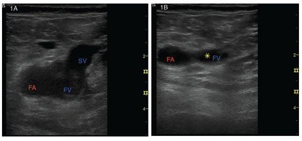
Intern Ultrasound of the Month: Superficial Thrombophlebitis — University  Hospitals Emergency Medicine Residency
Intern Ultrasound of the Month: Superficial Thrombophlebitis — University Hospitals Emergency Medicine Residency
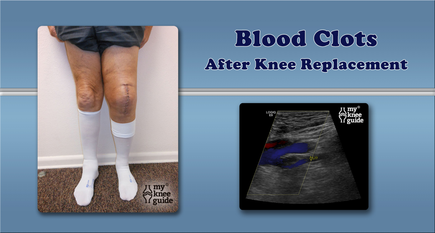
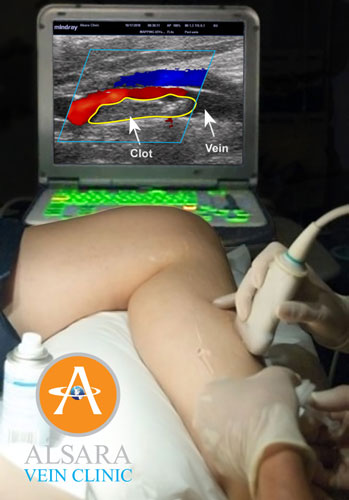
Blood Clots & Varicose Veins - 5X Higher Risk - Alsara Vein Clinic

Deep vein thrombosis | Radiology Reference Article | Radiopaedia.org

Deep Vein Thrombosis: Symptoms, Diagnosis and Treatment

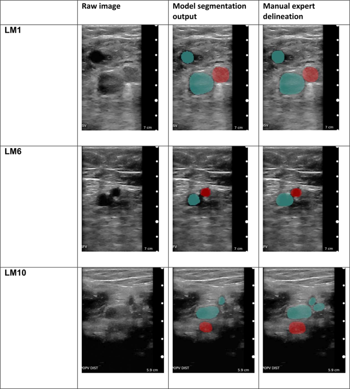
Non-invasive diagnosis of deep vein thrombosis from ultrasound imaging with  machine learning | npj Digital Medicine
Non-invasive diagnosis of deep vein thrombosis from ultrasound imaging with machine learning | npj Digital Medicine
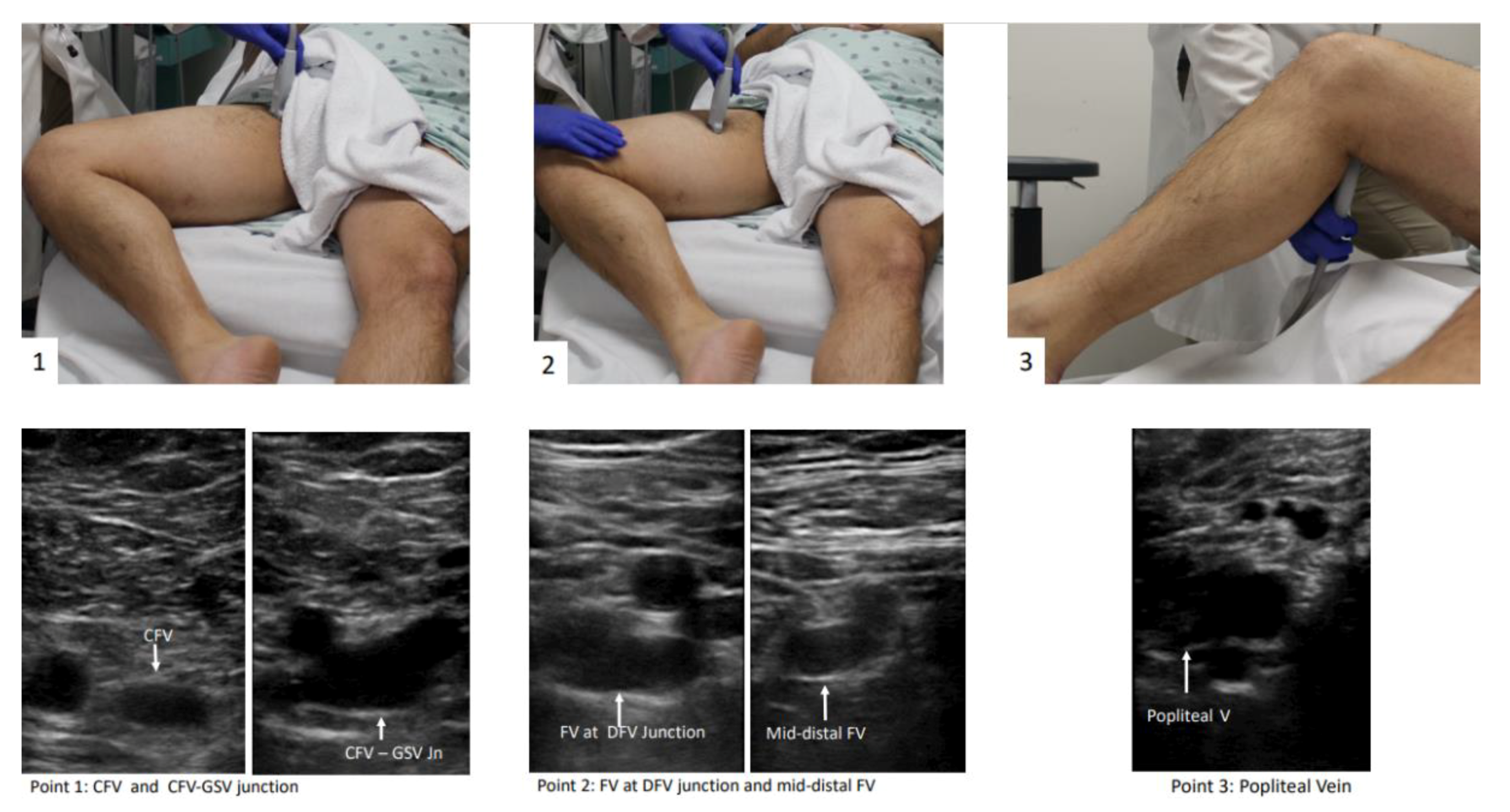
JCM | Free Full-Text | The Use of Point-of-Care Ultrasound (POCUS) in the  Diagnosis of Deep Vein Thrombosis
JCM | Free Full-Text | The Use of Point-of-Care Ultrasound (POCUS) in the Diagnosis of Deep Vein Thrombosis

Vein-Venous Doppler Ultrasound | pictureperfectultrasound

Massive DVT | Emory School of Medicine

What is a DVT Ultrasound?

POCUS - Lower Extremity Deep Venous Thrombosis (DVT) - Updated - YouTube

Deep Vein Thrombosis (DVT) - Vein Specialist of the Carolinas

DVT ultrasound - WikEM

Ultrasound Technique Breaks Down Blood Clots - Medical Design Briefs

Emergency Ultrasound: Deep Vein Thrombosis | MDedge Emergency Medicine

Ultrasound Technique Breaks Down Blood Clots - Medical Design Briefs

DVT Ultrasound: Procedure, Accuracy, Next Steps, and More

Doppler ultrasound showing blockage of the left superficial femoral... |  Download Scientific Diagram
Doppler ultrasound showing blockage of the left superficial femoral... | Download Scientific Diagram
Blood Clots: Also known as a DVT

Doppler ultrasound showing deep vein thrombosis - Stock Image - M175/0277 -  Science Photo Library
Doppler ultrasound showing deep vein thrombosis - Stock Image - M175/0277 - Science Photo Library

Harvard Trained Vein Doctor | Your Doctor Ordered Blood Clot Ultrasound Leg  Images – Now What?
Harvard Trained Vein Doctor | Your Doctor Ordered Blood Clot Ultrasound Leg Images – Now What?

Rheumatology Case Report: Deep Vein Thrombosis Detected by Point-of-Care  Ultrasound - The Rheumatologist
Rheumatology Case Report: Deep Vein Thrombosis Detected by Point-of-Care Ultrasound - The Rheumatologist

Deep Venous Thrombosis And Doppler Ultrasound - Dr. Sergio Mazzei | Wound  Care
Deep Venous Thrombosis And Doppler Ultrasound - Dr. Sergio Mazzei | Wound Care

Blood Clot in Leg on Ultrasound - YouTube

Ultrasonography of deep vein thrombosis - Wikipedia

Deep Vein Thrombosis Doppler Ultrasound Perth | Envision

Ultrasound for Lower Extremity Deep Venous Thrombosis | Circulation

Chronic recanalized deep vein thrombosis on ultrasound. Gray scale... |  Download Scientific Diagram
Chronic recanalized deep vein thrombosis on ultrasound. Gray scale... | Download Scientific Diagram